
News|Articles|May 31, 2025
When Medications Masquerade as Skin Disease
Listen
0:00 / 0:00
Key Takeaways
- Drug-induced skin reactions often present weeks to years after therapy initiation, complicating diagnosis and management.
- Autoimmune mimickers like dermatomyositis and subacute cutaneous lupus can be drug-induced, with statins and PPIs as notable triggers.
Scott Jackson, MD, explores complex drug-induced skin reactions, emphasizing diagnostic challenges and the importance of thorough medication reviews for effective treatment.
Advertisement
At the
Diagnostic Challenges and Delayed Onset
Jackson began by noting that many skin conditions commonly treated in dermatology clinics may actually be adverse drug reactions. Unlike immediate hypersensitivity reactions—such as penicillin rashes occurring within 48 hours—drug-induced dermatologic reactions often present weeks to months or even years after initiation of therapy. Furthermore, symptoms may persist long after the offending drug is discontinued, particularly in cases where the medication unmasks or triggers autoimmune processes, such as bullous pemphigoid.
Autoimmune Mimickers: Dermatomyositis and Subacute Cutaneous Lupus
Highlighting dermatomyositis, Jackson discussed statin-associated necrotizing autoimmune myopathy, a condition with cutaneous and systemic features mimicking classic dermatomyositis. He advocated for the use of HMGCR antibody assays when dermatomyositis-like presentations arise in statin-exposed patients.
Similarly, he emphasized the high rate of drug association in subacute cutaneous lupus erythematosus (SCLE), with up to one-third of cases being drug-induced. Proton pump inhibitors (PPIs) have emerged as notable triggers, likely due to their over-the-counter availability and intermittent use—both potential risk factors for sensitization.
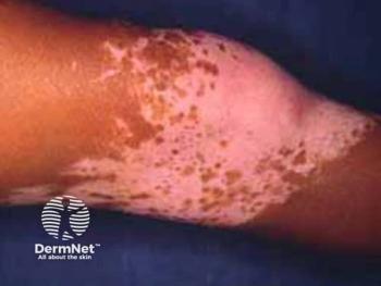
Fixed Drug Eruptions and Non-Pharmaceutical Triggers
Fixed drug eruptions (FDEs) remain a common presentation. Jackson drew attention to fluconazole, a frequently implicated agent in recent literature. Notably, he extended the discussion to non-pharmaceutical causes, such as fixed food eruptions and fixed alcohol eruptions, which can present similarly. Certain thermally stable antibiotics used in animal feed may even cause eruptions via food consumption—a diagnostic curveball for clinicians.
Dry Skin, Eczema, and Stasis Dermatitis
In elderly populations, xerosis and eczematous presentations can also be drug-induced. Jackson highlighted the role of statins and calcium channel blockers, such as amlodipine, particularly in cases of stasis dermatitis. He referenced studies that link amlodipine to both leg edema and eczematous reactions, reinforcing the need for a thorough medication history in evaluating chronic lower leg dermatitis.
Psoriasiform Dermatitis and Psoriasis Induction
Distinguishing drug-induced psoriasiform dermatitis from idiopathic psoriasis is another diagnostic challenge. Dr. Jackson cited abatacept, a rheumatoid arthritis biologic, as a common culprit. While statins may improve psoriasis, beta-blockers and certain weight-loss drugs (e.g., bupropion) have been implicated in exacerbating or inducing it. Additionally, antimalarials, particularly in predisposed patients, may provoke severe pustular psoriasis.
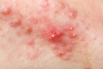
Acute Generalized Exanthematous Pustulosis (AGEP)
AGEP was discussed in the context of its clinical similarity to pustular psoriasis. A 2024 scoring system now aids clinicians in differentiating between the 2, with accurate identification remaining critical due to differences in management and prognosis.2
Bullous Pemphigoid and Biopsy Clues
Jackson concluded by revisiting bullous pemphigoid, noting that up to 22.5% of cases are drug-induced. Furosemide and dipeptidyl peptidase-4 (DPP-4) inhibitors remain key offenders. He also shared a diagnostic pearl for lichen planus-like eruptions: injecting lidocaine directly into the lesion prior to biopsy. The presence of a bulla post-injection may indicate a drug-induced lichenoid eruption rather than idiopathic lichen planus.
Clinical Takeaways
Jackson’s presentation underscored the importance of:
- Maintaining a high index of suspicion for drug-induced causes.
- Recognizing delayed reaction timelines.
- Considering non-pharmaceutical and food-related triggers.
- Utilizing updated diagnostic tools and antibody testing.
His lecture served as a reminder that dermatologic symptoms are often the tip of a pharmacologic iceberg—and that thorough, systematic medication reviews can dramatically alter the clinical trajectory for patients suffering from recalcitrant or atypical dermatoses.
By equipping clinicians with both new literature insights and practical diagnostic strategies, Jackson’s session reinforced the value of vigilance and curiosity in uncovering hidden iatrogenic causes of skin disease.
References
1. Jackson S. Drug induced skin disorders. Presentation at Fall Clinical PA/NP 2025 Conference; May 30-June 2, 2025; Orlando, Florida.
2. Yamanaka-Takaichi M, Watanabe M, Comfere NI, et al. Differentiating generalized pustular psoriasis from acute generalized exanthematous pustulosis. J Am Acad Dermatol. 2024;90(6):1289-1291. doi:10.1016/j.jaad.2024.01.080
Newsletter
Like what you’re reading? Subscribe to Dermatology Times for weekly updates on therapies, innovations, and real-world practice tips.
Advertisement
Related Content
Advertisement


Latest CME


Advertisement
Advertisement
Trending on Dermatology Times
1
FDA Accepts Galderma’s BLA Resubmission for Liquid Neuromodulator RelabotulinumtoxinA
2
AbbVie Files for Vitiligo Indication, Putting Systemic Therapy Under Regulatory Review
3
Icotrokinra Shows Superior Efficacy Over Advanced Oral Therapies in New Psoriasis Meta-Analysis
4
Introducing Dermatology Times NP/PA Connect
5