

News|Videos|June 28, 2025
Color as a Clinical Compass for Skin Cancer Detection
Author(s)Emma Andrus, Editor, Orit Markowitz, MD
Key Takeaways
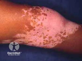
- Dr. Orit Markowitz advocates a color-first strategy for diagnosing skin cancer, enhancing early recognition of malignancy, especially in amelanotic melanomas.
- Prioritizing color assessment over pattern recognition simplifies the diagnostic process, aiding clinicians in diverse skin tones.
Orit Markowitz, MD, shares how a color-focused strategy helps spot early malignancies—even 2-mm lesions—across all skin tones.
Advertisement
At the 2025 Society of Dermatology Physician Associates Summer Dermatology Conference, Orit Markowitz, MD, founder and CEO of
In an interview, Markowitz explained how this approach improves diagnostic precision by keeping clinicians focused on what matters most: early recognition of malignancy, regardless of subtype or size.
“When we’re able to...decide whether something is benign or not, we’re already 10 steps ahead,” Markowitz said.
For many clinicians, particularly those newer to dermoscopy, pattern recognition can quickly become overwhelming. By leading with color assessment, both clinical and dermoscopic, Markowitz’s strategy offers a simplified path to diagnostic clarity. This is especially valuable when evaluating lesions in diverse skin tones, where pigmentation and morphology may vary.
“The other precedent for the color wheel approach was early amelanotic melanomas,” she said. “This is a topic that people are often a little bit anxious about—as they should be—because they’re difficult to diagnose, even on dermoscopy [and] noninvasive devices like confocal.”
Markowitz encourages clinicians to start with broad questions before diving into diagnostic complexity:
- What color is it clinically? Is it pink, pink-brown, or something else?
- What color is it dermoscopically?
- Does it raise suspicion before patterns even come into play?
“What this enables us to do is catch these lesions very, very early,” she said.
This early catch is critical, particularly for fast-progressing malignancies like amelanotic melanoma or early squamous cell carcinoma. In her lecture, Markowitz emphasized that recognizing certain basic color patterns without needing to precisely subtype the cancer right away can be enough to trigger action.
“Even if it’s 2 mm in diameter, it’s malignant,” she said. “I know it’s early malignant. I need to remove it.”
Instead of overprioritizing exact categorization at the outset, she advocates for a more practical decision-making framework.
“I’m not going to worry too much about what category of benign or malignant. I’m going to focus on the big picture. Does it need to come off?” she said.
That mindset, Markowitz explained, can improve both diagnostic speed and accuracy while minimizing unnecessary biopsies. It also makes the process more inclusive, as clinicians become trained to look at pigment and color more carefully across skin tones.
Make sure to keep up to date with the latest coverage from the conference and subscribe to Dermatology Times to receive daily email updates .
Newsletter
Like what you’re reading? Subscribe to Dermatology Times for weekly updates on therapies, innovations, and real-world practice tips.
Advertisement
Related Content
Advertisement


Latest CME


Advertisement
Advertisement
Trending on Dermatology Times
1
Icotrokinra Shows Superior Efficacy Over Advanced Oral Therapies in New Psoriasis Meta-Analysis
2
AbbVie Files for Vitiligo Indication, Putting Systemic Therapy Under Regulatory Review
3
Nutrafol Expands Portfolio with First and Only Hair Loss Supplement for Male Patients 50 and Older
4
Introducing Dermatology Times NP/PA Connect
5