
News|Articles|August 1, 2025
Patterns of Cutaneous Reactions to Nemolizumab in AD
Listen
0:00 / 0:00
Key Takeaways
- Nemolizumab is effective for pruritus in atopic dermatitis but may cause distinct cutaneous reactions in 40% of patients, often within the first three doses.
- Eruptions are typically non-pruritic and morphologically distinct from baseline AD exacerbations, with no significant correlation to disease severity or systemic inflammation markers.
The Japanese study found no association between cutaneous events and baseline EASI scores, eosinophil counts, or IgE levels.
Advertisement
Nemolizumab, a monoclonal antibody targeting the interleukin-31 receptor A (IL-31RA), has emerged as an important therapy for atopic dermatitis (AD) patients with moderate to severe pruritus.1 While its antipruritic efficacy is well established, new concerns have arisen regarding its association with distinct cutaneous manifestations.2-3 A recent Japanese multicenter retrospective study by Sasaki et al, published in The Journal of Dermatology, offers valuable clinical insights into the incidence, morphology, and potential risk factors for these adverse skin events in real-world settings.4
Study Overview
The study evaluated 219 patients aged ≥13 years with clinically diagnosed AD who received at least 1 dose of nemolizumab across 13 Japanese institutions between August 2022 and February 2024. Patients receiving fewer than 3 doses without any skin reactions were excluded to avoid misclassification of late-onset manifestations. Clinical features and laboratory markers were compared between patients with and without cutaneous adverse events.
The study was conducted using an international dosing regimen of 60 mg every 4 weeks, which differs from the FDA-approved US schedule of an initial 60 mg dose followed by 30 mg every 4 weeks. As a result, these findings may not be directly comparable to outcomes observed under the US-approved dosing.
Results
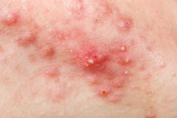
Researchers found cutaneous manifestations occurred in 88 patients (40.2%), typically within the first 3 doses. Erythema was the most prevalent eruption (69.3%), followed by coin-shaped red plaques with exudate (42.0%), dry/scaly lesions (29.5%), papules (21.6%), edema (10.2%), and vesicles (6.8%). Most eruptions (64.3%) were non-pruritic. Importantly, these manifestations were distinguished from exacerbations of baseline AD by clinical morphology and timing.
Two patients developed bullous pemphigoid, according to the study, confirmed by serologic and histopathologic testing, indicating a potential for nemolizumab to trigger autoimmune skin conditions in susceptible individuals.
The study found roughly 80% of cutaneous reactions emerged within the first 3 doses, with a significant drop in incidence after the sixth dose. The forearms, back, upper arms, and lower legs were the most frequently affected initial sites, suggesting a non-random distribution pattern that could aid early recognition.
Treatment and Outcomes
Despite the high incidence of reactions, 58% of affected patients continued nemolizumab therapy, typically managed with high-potency topical corticosteroids. The median duration of eruptions was 27 days for those who continued therapy and 33 days for those who discontinued, researchers stated, with no statistically significant difference. This suggests that stopping treatment does not necessarily lead to quicker resolution of symptoms.
The study found no significant associations between cutaneous manifestations and baseline disease severity (measured by EASI), eosinophil counts, serum IgE, or thymus and activation-regulated chemokine (TARC) levels.
These findings suggest that nemolizumab-induced eruptions may arise independently of known markers of AD severity or systemic inflammation. The absence of predictive biomarkers complicates efforts to stratify risk, emphasizing the need for close clinical observation early in treatment.
Pathophysiological Considerations
The study stated the distinct, often non-pruritic nature of these eruptions, coupled with their early onset and morphological variation, points toward a pharmacologic rather than disease-related mechanism. IL-31 is known to modulate neuroimmune interactions, and its blockade may disrupt regulatory feedback loops, possibly unleashing unchecked type 2 inflammation or unmasking latent autoimmunity.
Clinical Implications
For clinicians, awareness of these potential adverse effects is crucial for patient counseling and early management. Mild to moderate eruptions can typically be managed with intensified topical therapy, allowing for continued treatment. In severe cases, discontinuation may be necessary, but resolution should not be expected immediately due to nemolizumab's long half-life and prolonged pharmacodynamic effects.
Close monitoring during the initial dosing period—particularly up to the third dose—is advisable, according to researchers, and clinicians should be prepared to adjust treatment based on morphological patterns of eruptions rather than systemic biomarkers.
Conclusion
Nemolizumab is effective in controlling pruritus in AD, however some patients may experience distinct cutaneous reactions, often within the early phases of therapy. These reactions appear to be mechanistically independent of disease severity and immune status and can generally be managed with topical corticosteroids without discontinuing treatment. However, the absence of predictive markers highlights the importance of clinical vigilance and underscores the need for future research into the immunopathogenesis of these events.
References
- Igarashi A, Katsunuma T, Nagano Y, Komazaki H. Long-term (68 weeks) administration of nemolizumab in paediatric patients aged 6-12 years with atopic dermatitis with moderate-to-severe pruritus: efficacy and safety data from a phase III study. Br J Dermatol. 2025;192(5):837-844. doi:10.1093/bjd/ljae458
- Masuda T, Yonekura S, Kataoka K, et al. Psoriasis-like lesions in an atopic dermatitis patient possibly associated with nemolizumab treatment. J Dermatol. 2024;51(6):e193-e195. doi:10.1111/1346-8138.17085
- Katsuta M, Kamide R, Ishiuji Y, Nobeyama Y, Asahina A. Bullous pemphigoid that developed during nemolizumab treatment for atopic dermatitis: Two case reports. Acta Derm Venereol. 2024;104:adv40634. Published 2024 Sep 2. doi:10.2340/actadv.v104.40634
- Sasaki W, Saito R, Suzuki K, et al. Clinical characteristics and risk factors for cutaneous manifestations associated with nemolizumab in atopic dermatitis: A multicenter retrospective study in Japan. J Dermatol. Published online July 23, 2025. doi:10.1111/1346-8138.17877
Newsletter
Like what you’re reading? Subscribe to Dermatology Times for weekly updates on therapies, innovations, and real-world practice tips.
Advertisement
Related Content
Advertisement


Latest CME


Advertisement
Advertisement
Trending on Dermatology Times
1
Icotrokinra Shows Superior Efficacy Over Advanced Oral Therapies in New Psoriasis Meta-Analysis
2
AbbVie Files for Vitiligo Indication, Putting Systemic Therapy Under Regulatory Review
3
Nutrafol Expands Portfolio with First and Only Hair Loss Supplement for Male Patients 50 and Older
4
Introducing Dermatology Times NP/PA Connect
5