
News|Articles|May 2, 2025
OCT and D-OCT Imaging Reveal Distinct Vascular and Dermal Patterns in Skin Photoaging Phenotypes
Author(s)Emma Andrus, Editor
Listen
0:00 / 0:00
Key Takeaways
- OCT and D-OCT effectively identified distinct vascular and collagen patterns in atrophic and hypertrophic photoaging.
- Atrophic photoaging showed higher vascular density and collagen fragmentation compared to hypertrophic and control groups.
Collagen structure and vessel density vary by photoaging type, highlighting the value of multimodal imaging in guiding individualized skin care strategies.
Advertisement
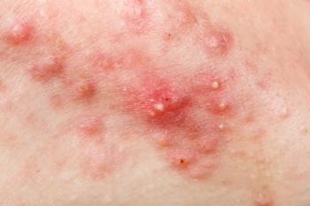
A recent study demonstrated the usefulness of optical coherence tomography (OCT) and dynamic OCT (D-OCT) for noninvasively identifying skin features associated with atrophic and hypertrophic photoaging.1
The study, published in the International Journal of Dermatology, reported that patients with atrophic photoaging exhibited D-OCT vessel assets and densities versus patients in either the control or hypertrophic photoaging cohorts.
Background and Methods
The present study was a post hoc analysis of data from a prospective imaging and genetic study conducted in Italy involving middle-aged Caucasian women. Participants were categorized into atrophic photoaging, hypertrophic photoaging, or control groups based on established clinical criteria.
Researchers conducted imaging using OCT and D-OCT on the cheek to assess vascular density and collagen structure and utilized additional data from standardized clinical photography and reflectance confocal microscopy2 to support and compare findings. Imaging assessments were conducted by multiple expert readers, and statistical correlations were analyzed to determine relationships between noninvasive imaging biomarkers and visible signs of aging.
Findings
The study evaluated 58 women categorized into atrophic photoaging, hypertrophic photoaging, and control groups. OCT and D-OCT imaging revealed distinct vascular and collagen patterns across the groups. While epidermal thickness and dermo-epidermal junction visibility were consistent among all participants, vascular differences were pronounced.
Specifically, the atrophic photoaging group exhibited significantly higher vascular density compared to both the hypertrophic photoaging and control groups (p=0.017), with 70.6% showing mild vascular presence and 17.6% showing moderate levels. These findings were complemented by fragmentation of collagen fibers, with moderate fragmentation being most common across all groups. However, patients with hypertrophic photoaging demonstrated slightly more severe collagen disorganization compared to controls.
Further correlations between imaging biomarkers highlighted how noninvasive assessments can reflect the underlying biological mechanisms of skin aging. In participants with atrophic photoaging, D-OCT-detected vessel density at both 300 and 500 μm depths was moderately to strongly associated with increased erythema (r=0.418 and r=0.450, respectively; p<0.05). Additionally, strong inter-layer correlations were observed between vessel densities at these 2 depths (r=0.790, p<0.01), as well as between collagen fiber disorganization and reflectance confocal microscopy (RCM) scores.
In participants with hypertrophic photoaging, skin aging presented a more fibrotic and thickened profile. Wrinkling correlated significantly with RCM collagen scores (r=0.477, p<0.05), while vessel density at 300 μm also aligned with areas of redness (r=0.526, p<0.05). RCM revealed curled and huddled collagen fibers—features that corresponded closely with epidermal irregularities and increased RCM collagen scores.
Conclusions
The study may have been limited by factors such as its retrospective design and limited sample size, as well as potential subject selection bias, including leaning primarily toward toward female participants.
"This study contributes to our current knowledge of different morphologic details, revealing different response pathways to photo exposure," wrote study authors Guida et al. "In the era of personalized medicine, optimal treatment should focus on the morphologic differences in skin photoaging patterns."
Moving forward, larger, prospective studies including are needed to validate these preliminary findings and establish clinical thresholds for noninvasive imaging metrics.
References
- Guida S, Ciardo S, Galadari H, et al. Correlating optical coherence tomography and other noninvasive imaging features with atrophic and hypertrophic skin photoaging. Int J Dermatol. Published online April 18, 2025.
doi:10.1111/ijd.17799 - Levine A, Markowitz O. Introduction to reflectance confocal microscopy and its use in clinical practice. JAAD Case Rep. 2018;4(10):1014-1023. Published 2018 Nov 10.
doi:10.1016/j.jdcr.2018.09.019
Newsletter
Like what you’re reading? Subscribe to Dermatology Times for weekly updates on therapies, innovations, and real-world practice tips.
Advertisement
Related Content
Advertisement


Latest CME


Advertisement
Advertisement
Trending on Dermatology Times
1
Icotrokinra Shows Superior Efficacy Over Advanced Oral Therapies in New Psoriasis Meta-Analysis
2
AbbVie Files for Vitiligo Indication, Putting Systemic Therapy Under Regulatory Review
3
Nutrafol Expands Portfolio with First and Only Hair Loss Supplement for Male Patients 50 and Older
4
Introducing Dermatology Times NP/PA Connect
5