
News|Articles|July 16, 2025
Journal Digest: July 16, 2025
Author(s)Emma Andrus, Editor
Listen
0:00 / 0:00
Key Takeaways
- Expert guidelines stress structured safety protocols to prevent ocular complications during laser procedures near the eyes.
- Radiation dermatitis management requires tailored strategies, with aggressive intervention for internal cancers and restraint for nonmelanoma skin cancers.
This review of the latest dermatologic studies includes insights into ocular safety during laser procedures, radiation dermatitis prevention and management, and more.
Advertisement
Journal of Cosmetic Dermatology: Expert Consensus on Ocular Safety During Laser Procedures: A Practical Guide to Eye Safety by a Panel of Dermatologist Laser Surgeons and Ophthalmologists
A recent expert consensus offered comprehensive guidelines to minimize ocular risks during laser procedures near the eyes. The panel, composed of dermatologic laser surgeons and ophthalmologists, emphasized that serious complications such as corneal burns, dry eye, cataracts, and blindness can be avoided through structured safety protocols. Their recommendations included preoperative screening for ocular and systemic risk factors, intraoperative use of lubricated stainless steel eye shields with real-time monitoring, and tailored postoperative care including sterile drops and patient education.1
Dermatological Reviews: Radiation Dermatitis: A Comparative Review of Prevention and Management
A recent review examined the differing approaches to managing radiation dermatitis in patients undergoing radiation therapy for internal malignancies versus nonmelanoma skin cancers. The authors detailed the pathophysiology of radiation dermatitis, marked by inflammation, barrier dysfunction, and cellular damage, and discussed risk factors and clinical manifestations of both acute and chronic radiation dermatitis. They review highlighted the importance of tailoring radiation dermatitis prevention and treatment strategies to the clinical context, prioritizing aggressive intervention for internal cancers and a more restrained approach during active superficial radiation therapy for nonmelanoma skin cancers.2
Skin Research and Technology: Seborrheic Keratosis-Like Melanoma: Novel Insights Into Clinical, Dermoscopic, and Reflectance Confocal Microscopy Diagnosis of an Atypical Melanoma Variant
A recent retrospective study investigated the clinical, dermoscopic, and reflectance confocal microscopy (RCM) features of seborrheic keratosis-like melanoma (SKLM). Among 60 histologically confirmed SKLM cases, the majority were superficial spreading melanomas, frequently located on the trunk and most often affecting men under 50. Dermoscopic analysis revealed that all lesions exhibited at least 1 melanoma-specific criterion, justifying excision. RCM revealed consistent melanoma-associated features such as irregular epidermal patterns, atypical nests, dendritic cells, and dermal inflammation.3
JAMA Dermatology: Risk Factors for Incident Nevus-Associated vs De Novo Invasive Melanoma
A recent prospective cohort study from the QSkin Study examined risk factor differences between nevus-associated melanom and de novo melanoma, analyzing data from 859 individuals over 11.4 years. The study found that high nevus density and elevated polygenic risk scores for melanoma were more strongly associated with NAM than with de novo melanoma. While overall risk profiles were similar across sexes, NAMs were more commonly located on the trunk in males and on the limbs in females.4
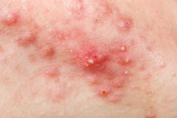
Lasers in Surgery and Medicine: Treatment of Cutaneous Collagenous Vasculopathy With Pulsed Dye Laser
A recent case report outlined the case of a 40-year-old woman with extensive cutaneous collagenous vasculopathy (CCV) involving her extremities and trunk who was successfully treated with pulsed dye laser (PDL) therapy over 14 months. The treatment, using a 595-nm PDL, led to significant and sustained improvement in telangiectasias, erythema, discomfort, and overall cosmetic appearance. Adjustments in pulse duration and spot size optimized tolerability and efficacy. According to authors, this case adds to the limited literature on PDL for CCV and supports its safe and effective use as a therapeutic option for this rare vascular condition with challenging clinical management.5
References
- Kesty K, Kesty C, Benson J, Kesty K, Kesty C. Expert consensus on ocular safety during laser procedures: a practical guide to eye safety by a panel of dermatologist laser surgeons and ophthalmologists. J Cosmet Dermatol. Published online July 10, 2025.
doi:10.1111/jocd.70339 - Chaudry A, Vanaria RJ, Nestor MS. Radiation dermatitis: a comparative review of prevention and management. Dermatol Rev. Published online July 11, 2025.
doi:10.1002/der2.70043 - Venturi F, Cedirian S, Veronesi G, Scotti B, Baraldi C, Dika E. Seborrheic keratosis-like melanoma: novel insights into clinical, dermoscopic, and reflectance confocal microscopy diagnosis of an atypical melanoma variant. Skin Res Technol. Published online July 11, 2025.
doi:10.1111/srt.70206 - Olsen CM, Shalit MM, Pandeya N, et al. Risk factors for incident nevus-associated vs de novo invasive melanoma. JAMA Dermatol. Published online July 9, 2025.
doi:10.1001/jamadermatol.2025.2004 - Hanania HL, Camacho-Hubbard I, Nawas ZY. Treatment of cutaneous collagenous vasculopathy with pulsed dye laser. Lasers Surg Med. Published online July 15, 2025.
doi:10.1002/lsm.70048
What new studies have you been involved with or authored? Share with us by emailing
Newsletter
Like what you’re reading? Subscribe to Dermatology Times for weekly updates on therapies, innovations, and real-world practice tips.
Advertisement
Related Content
Advertisement


Latest CME


Advertisement
Advertisement
Trending on Dermatology Times
1
FDA Accepts Galderma’s BLA Resubmission for Liquid Neuromodulator RelabotulinumtoxinA
2
AbbVie Files for Vitiligo Indication, Putting Systemic Therapy Under Regulatory Review
3
Icotrokinra Shows Superior Efficacy Over Advanced Oral Therapies in New Psoriasis Meta-Analysis
4
Introducing Dermatology Times NP/PA Connect
5