
News|Articles|July 15, 2025
Transcriptomics Reveal Core ACD Immune Pathways
Listen
0:00 / 0:00
Key Takeaways
- Transcriptomic analysis of severe patch test reactions identified over 800 commonly upregulated genes across allergens, highlighting shared innate and adaptive immune pathways.
- Allergen-specific molecular patterns emerged, with nickel and 2-HEMA showing distinct DEG sets, suggesting dual inflammatory-irritant profiles and unique immune activations.
New insights reveal shared and allergen-specific immune responses in allergic contact dermatitis.
Advertisement
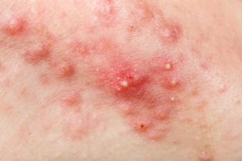
While patch testing remains the cornerstone of diagnosis for allergic contact dermatitis (ACD), the immunologic events driving elicitation—the phase responsible for symptom onset—are incompletely understood.1 A recent study by Pesqué et al published in Allergy, provides new clinical and molecular insights into this process by conducting a comprehensive transcriptomic analysis of strong and extreme patch test reactions in non-atopic individuals.2
Methods and Materials
The study examined 40 severe (2+ or 3+) patch test reactions to 4 chemically distinct allergens: nickel, 2-hydroxyethylmethacrylate (2-HEMA), methylisothiazolinone (MIT), and formaldehyde. Only non-atopic individuals, verified through stringent diagnostic criteria and dermatological assessments, were included to minimize confounding from comorbid atopic dermatitis, which is known to alter ACD immune profiles. Nineteen petrolatum-exposed controls were also analyzed.
RNA sequencing of 3 mm punch biopsies collected 96 hours post-patch test revealed a robust set of differentially expressed genes (DEGs), with over 800 genes commonly upregulated across all allergens compared to controls. Key shared pathways were dominated by both innate and adaptive immune responses. Top DEGs included chemokines (e.g., CXCL10, CCL17), cytokine regulators (e.g., IL13, IL23A), and markers of dendritic cell activation and T-cell signaling.
Results
Although a strong common “ACD transcriptomic signature” was identified, researchers stated notable allergen-specific molecular patterns also emerged. Nickel and 2-HEMA reactions in particular exhibited distinct sets of DEGs. Nickel-specific genes were associated with cell cycle and innate immune responses—some overlapping with previously described irritant contact dermatitis pathways—raising the possibility of a dual inflammatory-irritant profile. In contrast, 2-HEMA uniquely activated B-cell receptor and antigen processing pathways, alongside markers of type 2 (e.g., CCL11) and type 1 (e.g., IL18R1) immunity.
Researchers noted allergens uniformly elicited mixed T-helper cell responses. Type 1/Th1 pathways were dominant, researchers noted, with strong IFN-γ–related signatures. Type 3/Th17 responses also featured prominently, reinforcing recent findings of their role in ACD pathology. Type 2/Th2 signals were present but less prominent. These findings contrast with previous research that suggested greater Th2 involvement, particularly in atopic individuals or fragrance allergens, highlighting the impact of comorbidities and allergen type.
Interestingly, partially shared DEGs between allergen pairs further delineated subgroup-specific immunologic features, such as IL6R and LOXL3 in 2-HEMA and MIT combinations, and Th2-biased markers in certain mixtures. Researchers said these partially shared genes contribute to the complexity of immune polarization in ACD.
Downregulated genes across all allergens were primarily associated with epidermal structural components (e.g., FLG2, LORICRIN), indicating impaired skin barrier homeostasis during elicitation. This aligns with the visible spongiotic features noted histologically in all reactions and supports the hypothesis that inflammation-driven barrier dysfunction is intrinsic to ACD pathogenesis.
Conclusion
The study is notable for its rigorous exclusion of atopic dermatitis and its focus on strong-to-extreme ACD reactions, which may explain the amplified core inflammatory transcriptomic profile observed. However, limitations include the small cohort size, and the restricted number of allergens studied. Additional research across broader patient populations and allergen classes is needed to validate these findings and explore clinical implications, particularly for biomarker development or therapeutic modulation.
This research highlights a dual pattern of shared and allergen-specific immune responses in ACD elicitation, dominated by type 1 and type 3 immunity in non-atopic individuals. Researchers behind the study hope these insights advance understanding of ACD pathophysiology and provide a foundation for more precise diagnostic and therapeutic strategies.
References
- Dhingra N, Shemer A, Correa da Rosa J, et al. Molecular profiling of contact dermatitis skin identifies allergen-dependent differences in immune response. J Allergy Clin Immunol. 2014;134(2):362-372. doi:10.1016/j.jaci.2014.03.009
- Pesqué D, Andrades E, Berenguer-Molins P, et al. Transcriptomic analysis of allergic patch test reactions in non-atopic patients: A comparative study across multiple allergens. Allergy. Published online July 9, 2025. doi:10.1111/all.16642
Newsletter
Like what you’re reading? Subscribe to Dermatology Times for weekly updates on therapies, innovations, and real-world practice tips.
Advertisement
Related Content
Advertisement


Latest CME


Advertisement
Advertisement
Trending on Dermatology Times
1
AbbVie Files for Vitiligo Indication, Putting Systemic Therapy Under Regulatory Review
2
FDA Accepts Galderma’s BLA Resubmission for Liquid Neuromodulator RelabotulinumtoxinA
3
Icotrokinra Shows Superior Efficacy Over Advanced Oral Therapies in New Psoriasis Meta-Analysis
4
Nutrafol Expands Portfolio with First and Only Hair Loss Supplement for Male Patients 50 and Older
5