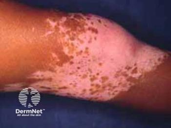
Review Highlights Treg Cell Impairment as Key Factor in Vitiligo
Key Takeaways
- Treg cell deficiencies, both numerical and functional, are significantly linked to vitiligo, suggesting their role as therapeutic targets.
- Subtle immune imbalances, particularly involving CD8+ cytotoxic T cells, are central to vitiligo pathogenesis.
A systematic review reveals significant Treg cell deficiencies in vitiligo, highlighting their role as therapeutic targets for improved treatment strategies.
A recent systematic review and meta-analysis of 21 case-control studies highlighted that both numerical and functional deficiencies in regulatory T (Treg) cells are significantly associated with vitiligo, with reviewers placing an emphasis on their role as therapeutic targets in the disease’s pathogenesis.1
Authors of the review, published in the International Journal of Dermatology, observed that while overt systemic inflammation is not characteristic of vitiligo, subtle immune imbalances, particularly involving CD8+ cytotoxic T cells and insufficient regulatory control, may continue to be central to understanding disease onset and progression.
Background and Methods
Clinical research in vitiligo already points to functional disturbance of Tregs in patients with vitiligo. A 2022 study published in the Journal of Autoimmunity reported that in vitiligo, Tregs lose their suppressive function and adopt a dysfunctional Th1-like phenotype.2 This leads toCD8+ T cell–mediated melanocyte destruction, the hallmark of vitiligo pathogenesis.
The present meta-analysis, registered with PROSPERO and conducted according to PRISMA guidelines, examined the frequency and suppressive capacity of Treg cells in individuals with vitiligo compared with healthy controls.
Inclusion criteria limited studies to those using fluorescence-activated cell sorting or equivalent methodologies to define Treg cells, and outcomes included cell counts, cytokine profiles (IL-10, TGF-β), and FOXP3 expression.
Researchers analyzed data from 21 studies (including 1016 cases of vitiligo and data from 846 healthy controls), with meta-analyses performed for blood-based Treg metrics and narrative synthesis for skin-based assessments.
Findings
Reviewers noted that a prior meta-analysis of 11 studies (518 patients with vitiligo and 437 controls) revealed a statistically significant decrease in circulating Treg cell frequency in vitiligo. Meta-regression showed no significant correlation between disease activity status (active vs stable) and Treg count reductions.
While skin-based data were less consistent, most studies reported reduced Treg cell numbers in lesional and perilesional skin compared to healthy controls. Some variability existed, with 2 studies showing increased or unchanged levels in certain anatomical regions. Regardless, most findings pointed toward an insufficient Treg presence in areas of immune activity, suggesting impaired immune regulation at the site of melanocyte loss.
Seven studies evaluating Treg suppressive function using co-culture and CFSE-based assays showed that Treg cells from patients with vitiligo exhibited a reduced capacity to suppress effector T cells, especially CD8+ subsets. The overall suppressive deficit was significant, and suppression of CD8+ T cells was particularly impaired, while suppression of CD4+ T cells showed no significant difference.
Three studies assessing IL-10 levels reported significantly decreased serum concentrations in patients with vitiligo. TGF-β levels, assessed in 5 studies, showed a nonsignificant trend toward reduction.
Meta-analysis also confirmed elevated serum levels of IL-17 and IL-22: cytokines typically associated with Th17 cells and chronic inflammation. IL-17 levels were increased, as were IL-22 levels, though their role in vitiligo remained debated.
The transcription factor FOXP3, a regulator of Treg identity and function, was consistently found to be downregulated in Treg cells from the blood of patients with vitiligo across multiple studies. Skin-based FOXP3 data were more variable, but most studies reported reduced expression in lesional tissue, aligning with diminished local immune tolerance.
Conclusions and Future Directions
The findings of this meta-analysis add support to the hypothesis that vitiligo involves not only autoreactive CD8+ T cell aggression but also a failure in regulatory T cell control.
Heterogeneity in study design, clinical phenotypes, sample handling, and control selection may complicate interpretation of the review’s findings. Skin-based studies in particular lacked standardization in biopsy location and disease phase, limiting cross-comparability. To address these concerns, review authors employed random-effects models and sensitivity analyses.
“Taken together, a quantitative reduction and functional impairments of Treg cells emerge as a core immunopathological feature in vitiligo patients,” wrote review authors Lerner et al. “A better understanding of the role of Treg cells in vitiligo pathogenesis will pave the way for the development of innovative and targeted therapeutic strategies to effectively treat patients with this condition.”
Further studies with phenotyping, standardized immunologic assays, and controlled longitudinal designs are needed to validate Treg cells as both biomarkers and therapeutic targets in vitiligo.
References
- Lerner G, Nikolaou M, Stoffel C, et al. Regulatory T cell dysregulation in vitiligo: a meta-analysis and systematic review of immune mechanisms and therapeutic perspectives. Int J Dermatol. Published online July 14, 2025. doi:10.1111/ijd.17959
- Chen J, Wang X, Cui T, et al. Th1-like Treg in vitiligo: An incompetent regulator in immune tolerance. J Autoimmun. 2022;131:102859. doi:10.1016/j.jaut.2022.102859
Newsletter
Like what you’re reading? Subscribe to Dermatology Times for weekly updates on therapies, innovations, and real-world practice tips.


