
News|Articles|April 10, 2025
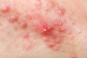
Microneedles Offer a New Hope for Nodular Acne Treatment
Listen
0:00 / 0:00
Key Takeaways
- Dissolvable microneedles enhance clindamycin delivery to sebaceous glands, improving nodular acne treatment efficacy and reducing systemic exposure.
- PVA-based microneedles exhibit optimal mechanical properties, penetration capacity, and rapid drug release, supporting their therapeutic potential.
Microneedles successfully penetrated simulated skin barriers, suggesting their effectiveness in reaching sebaceous glands.
Advertisement
Nodular acne can present challenges for conventional therapies due to limited drug penetration and systemic adverse effects.1 In response, Sangnim et al. explored the development of dissolvable microneedles (MNs) as a targeted delivery system for clindamycin, a topical antibiotic commonly used in acne treatment.2
The study aimed to overcome limitations of traditional topical and systemic treatments by using microneedle-based delivery to localize clindamycin administration directly into the sebaceous glands, the typical site of nodular acne pathogenesis. Conventional clindamycin formulations often fail to reach therapeutic concentrations in these deeper skin layers, whereas oral isotretinoin—effective for severe cases—is associated with significant adverse effects. The MNs were hypothesized to bypass the stratum corneum and allow precise, localized drug delivery, thereby enhancing efficacy while reducing systemic exposure.
Methods
The researchers employed chitosan, polyvinylpyrrolidone (PVP), and polyvinyl alcohol (PVA) to fabricate microneedles through a mold-based technique supported by stereolithographic 3D printing. Designs included cone-shaped and square pyramid-shaped microneedles with lengths ranging from 2000 to 3000 μm. Among these, PVA-based microneedles proved most effective due to their robust mechanical properties and ease of mold removal. Further enhancement was achieved by cross-linking the PVA MNs in sodium sulfate, which improved structural rigidity and ensured cleaner detachment from molds.
The microneedles were evaluated based on morphology, mechanical strength, penetration capacity, and drug release behavior. Scanning Electron Microscopy (SEM) revealed that PVA microneedles maintained sharp, layered, and uniform structures with solid adhesion to the PEG10000 base, used to support application and stability. The morphology was consistent across different angles, and the integration with the PEG base enhanced the integrity and user handling of the patches.
Mechanical analysis using a texture analyzer demonstrated that the cone-shaped PVA microneedles, particularly those measuring 2500 μm, exhibited the highest breaking force. This structural resilience was attributed to the even distribution of stress in the conical design. Moreover, shorter microneedles were mechanically superior to longer ones, likely due to reduced susceptibility to buckling and bending forces, which could otherwise lead to breakage.
Results
In vitro penetration testing using Parafilm—a synthetic skin analog—showed that the microneedles successfully penetrated 4 layers, representing a depth of approximately 500 μm, and in some instances reached 8 layers (approximately 1 mm). These results suggest sufficient penetration to access sebaceous glands located in the dermis, making the approach viable for treating deep-seated nodular acne lesions. SEM analysis of the microneedles post-penetration indicated a potential for controlled detachment from the base, which could facilitate localized drug delivery while leaving the active component embedded in the skin.
Drug release testing revealed a rapid and complete release of clindamycin within 3 minutes in phosphate buffer solution, indicating a desirable fast-dissolving profile. This release pattern supports efficient therapeutic action and minimizes the duration of skin contact, which can help reduce the risk of irritation. The rapid dissolution aligns well with the concentration-dependent antimicrobial activity of clindamycin and the clinical need for immediate therapeutic response.
Conclusion
Overall, the study demonstrates that PVA-based dissolvable microneedles offer a promising platform for the localized treatment of nodular acne. Their optimized mechanical properties, effective penetration, and rapid drug release underscore their potential to improve upon current topical and oral therapies. Nevertheless, further investigations using ex vivo and in vivo models are needed to validate these findings in clinically relevant conditions. Researchers suggested future research should also address the scalability of the fabrication process and explore regulatory pathways for eventual clinical implementation.
References
- Vasam M, Korutla S, Bohara RA. Acne vulgaris: A review of the pathophysiology, treatment, and recent nanotechnology based advances. BiochemBiophys Rep. 2023;36:101578. Published 2023 Nov 23. doi:10.1016/j.bbrep.2023.101578
- Sangnim T, Panpipat C, Chonsupawan S, et al. Development of clindamycin-loaded microneedles for the treatment of nodular acne: A novel therapeutic approach. Dermatology Research and Practice. 2025. doi:10.1155/drp/2138049
Newsletter
Like what you’re reading? Subscribe to Dermatology Times for weekly updates on therapies, innovations, and real-world practice tips.
Advertisement
Related Content
Advertisement


Latest CME


Advertisement
Advertisement
Trending on Dermatology Times
1
Icotrokinra Shows Superior Efficacy Over Advanced Oral Therapies in New Psoriasis Meta-Analysis
2
AbbVie Files for Vitiligo Indication, Putting Systemic Therapy Under Regulatory Review
3
Nutrafol Expands Portfolio with First and Only Hair Loss Supplement for Male Patients 50 and Older
4
Introducing Dermatology Times NP/PA Connect
5