
News|Articles|April 16, 2025
Journal Digest: April 16
Author(s)Emma Andrus, Editor
Listen
0:00 / 0:00
Key Takeaways
- Dermatologic associations in Crohn's disease patients emphasize the need for collaboration between dermatology and gastroenterology.
- Tinea pedis prevalence in children varies globally, with Trichophyton rubrum as a leading cause.
This review of the latest dermatologic studies includes insights into dermatologic associations in patients with Crohn disease, the prevalence of tinea pedis in pediatric populations, and more.
Advertisement
JEADV Clinical Practice: Exploring the Cutaneous Associations of Crohn's Disease: A Retrospective Study of 108 Patients
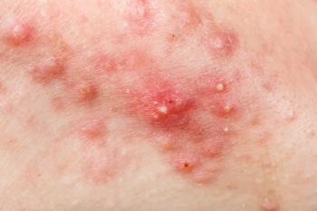
A recent retrospective chart review examined dermatologic associations in patients with Crohn disease seen at Wake Forest Baptist Dermatology between 2010 and 2024. Researchers analyzed 108 patient records, with statistical comparisons made using χ² and 2-tailed t-tests. The majority of patients were female (54.6%) and White (80.6%). Over half (57.4%) had at least 1 associated dermatologic diagnosis. The study proposes a 5-tiered grouping system to describe these associations and emphasizes the need for multidisciplinary collaboration between dermatology and gastroenterology.1
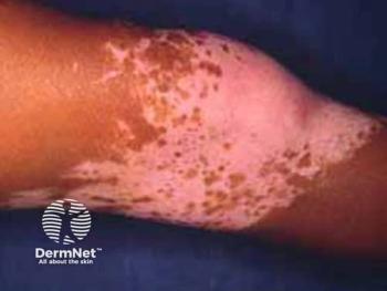
Pediatric Dermatology: Systematic Review of the Prevalence of Tinea Pedis in Children
A recent systematic review investigated the prevalence of tinea pedis in pediatric populations. Researchers analyzed 29 studies published through October 10, 2024, each including at least 100 children aged 0–19 years. Reported prevalence rates ranged from 0.03% to 15.6%, with clinical examination, microscopy, and culture identified as the most common diagnostic methods. Dermatophytes, particularly Trichophyton rubrum, were the leading etiological agents. The review highlights global variability in prevalence and emphasizes the importance of clinician awareness when diagnosing tinea pedis in children.2
Health Science Reports: The Efficacy of Platelet-Rich Plasma in Facial Lichen Planus Pigmentosus: A Prospective Pilot Study
A recent pilot study explored the use of platelet-rich plasma (PRP) as a novel treatment for lichen planus pigmentosus (LPP). Twelve patients received 3 sessions of intradermal PRP injections over 4 weeks, with follow-up visits extending to week 12. Researchers observed a significant reduction in melanin index and improvement in physician global assessment scores and patient satisfaction. By week 12, 41.7% of patients reported more than 50% improvement in their lesions.3
Journal der Deutschen Dermatologischen Gesellschaft: Combination of LED illumination and daylight photodynamic therapy for the treatment of actinic keratosis in solid organ transplant recipients: a prospective, randomized, comparative, intra-patient study
A recent randomized intra-patient study evaluated the effectiveness of combining daylight photodynamic therapy (DL-PDT) with LED illumination versus DL-PDT alone in treating actinic keratosis (AK) among solid organ transplant recipients. Conducted at the University of Navarra Clinic, the study involved 13 patients with 2 comparable facial or scalp areas treated with DL-PDT, 1 of which also received additional LED illumination. After 12 weeks, the combination therapy showed a significantly greater reduction in AKs (79.55%) compared to DL-PDT alone (65.43%). Findings suggest enhanced efficacy with the combined approach in this high-risk population.4
Journal of Cutaneous Pathology: Histopathologic Evaluation of Density and Depth of the Lymphoid Infiltrate in Clinically Defined Patches and Plaques in Early Stage Mycosis Fungoides
A recent study explored whether histopathologic criteria, specifically, the extent and depth of lymphocytic infiltrates, could reliably distinguish between patches and plaques in early-stage mycosis fungoides. Involving 100 biopsy samples, the study categorized infiltrates into 4 histopathologic levels and compared them with clinical classifications. Results showed that minimal-to-mild infiltrates (category 1) strongly correlated with patches (88%), but deeper infiltrates (category 4) were found in both patches (55%) and plaques (45%). This suggests that infiltrate depth alone is insufficient for differentiating lesion types.5
References
- Oscherwitz ME, Ricardo JW, Vescovacci NM, Kohrs AB, Jorizzo JL. Exploring the cutaneous associations of Crohn's disease: A retrospective study of 108 patients. JEADV Clin Pract. 2025;1(2):e70036.
doi:10.1002/jvc2.70036 - Stenderup JEB, Goandal NF, Saunte DML. Systematic review of the prevalence of tinea pedis in children. Pediatr Dermatol. 2025;42(2):e15947.
doi:10.1111/pde.15947 - Kiatsurayanon C, Deeudomwongsa P, Pituvong P, Sajjachareonpong P. The efficacy of platelet-rich plasma in facial lichen planus pigmentosus: A prospective pilot study. Health Sci Rep. 2025;8(4):e70455.
doi:10.1002/hsr2.70455 - Oteiza-Rius I, Morelló-Vicente A, Aguado-Gil L, et al. Combination of LED illumination and daylight photodynamic therapy for the treatment of actinic keratosis in solid organ transplant recipients: a prospective, randomized, comparative, intra-patient study. J Dtsch Dermatol Ges. 2025;23(4):e15665.
doi:10.1111/ddg.15665 - Kersten JM, Ottevanger R, Doeleman T, et al. Histopathologic evaluation of density and depth of the lymphoid infiltrate in clinically defined patches and plaques in early stage mycosis fungoides. J Cutan Pathol. 2025;52(4):e14810.
doi:10.1111/cup.14810
What new studies have you been involved with or authored? Share with us by emailing
Newsletter
Like what you’re reading? Subscribe to Dermatology Times for weekly updates on therapies, innovations, and real-world practice tips.
Advertisement
Related Content
Advertisement


Latest CME


Advertisement
Advertisement
Trending on Dermatology Times
1
Icotrokinra Shows Superior Efficacy Over Advanced Oral Therapies in New Psoriasis Meta-Analysis
2
AbbVie Files for Vitiligo Indication, Putting Systemic Therapy Under Regulatory Review
3
Nutrafol Expands Portfolio with First and Only Hair Loss Supplement for Male Patients 50 and Older
4
Introducing Dermatology Times NP/PA Connect
5