Five Clinical Subtypes Identified in Vitiligo Cohort
Key Takeaways
- Personalized medicine in vitiligo treatment is advancing, with topical JAK inhibitors like ruxolitinib approved in the US and Europe.
- A study identified five distinct clinical phenotypes in non-segmental vitiligo, aiding in tailored management strategies.
Recent research outlined 5 subtypes of vitiligo that can be used to guide therapeutic choices.
Although the precise etiology of vitiligo remains complex, involving a combination of genetic predisposition and immune dysregulation, recent therapeutic advancements have shifted focus toward personalized medicine. Most notably, topical Janus kinase inhibitors such as ruxolitinib have received approval in the US and Europe, signaling a new era in targeted therapy.1 However, clinical heterogeneity among patients continues to challenge standard classification and treatment.
A recent single-center observational study conducted at the University of Bordeaux aimed to address this issue by identifying distinct clinical phenotypes among individuals with nonsegmental vitiligo.2 Using a combination of factor analysis of mixed data (FAMD) and consensus clustering techniques, the study stratified 399 patients into 5 reproducible and clinically meaningful clusters. This approach moves beyond the conventional classification of vitiligo, which typically centers on lesion distribution alone.
Study Design and Methodology
Patients included in the study had a confirmed diagnosis of nonsegmental vitiligo and had not received any systemic or topical treatments within the prior year. Comprehensive clinical data, including lesion localization, disease activity signs (eg, trichrome and confetti lesions), family history, autoimmune comorbidities, and Koebner phenomenon in vitiligo scores (K-VSCOR), were systematically collected using a standardized physician-administered questionnaire.
The data set was analyzed using FAMD to reduce dimensionality and account for both continuous and categorical variables. K-means consensus clustering was then applied to identify stable phenotypic groups.
Identified Patient Clusters
Highly Active Vitiligo (12.5%)
This group exhibited the most clinical activity, including high frequencies of trichrome (96%) and confetti lesions (80%), along with trauma-associated depigmentation (Koebner type 2B). Patients had moderate surface involvement (mean Vitiligo Extent Score [VES]: 9.27) and commonly experienced pruritus. Systemic therapies, including oral steroid minipulses (60%) and narrowband UVB (60%), were frequently prescribed. This cluster is of particular interest because of its elevated risk of rapid progression, warranting early therapeutic intervention.
Mild Vitiligo (31.8%)
Characterized by limited disease extent (mean VES: 1.49) and minimal signs of activity, this group primarily had facial and acral lesions. Topical treatments were predominant, but more than half of these patients (52%) were lost to follow-up, possibly reflecting low perceived disease burden or treatment dissatisfaction.
Extensive Vitiligo (12.3%)
Patients in this group had widespread disease (mean VES: 32.4), prolonged duration (mean: 24.9 years), and the highest incidence of leukotrichia (26.5%). Nearly half received systemic therapy, but treatment response is often limited because of the chronicity and depigmented hair. This cluster exemplifies treatment resistance and highlights the need for more robust therapeutic strategies.
Moderate to Severe Type 2A Koebner Vitiligo (13.3%)
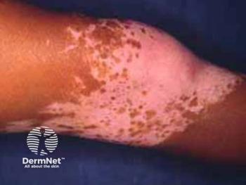
With lesions largely confined to friction-prone areas (eg, elbows, knees, waistline), this cluster shared pathophysiological similarities with psoriasis. Despite moderate disease extent (mean VES: 7.04), patients displayed low disease activity, favoring topical treatments with supplemental phototherapy in select cases.
Mild Type 2A Koebner Vitiligo (30%)
This cluster was the largest and was defined by localized lesions in classic Koebner sites and acral regions, with low disease activity and moderate K-VSCOR values. Notably, this group showed the highest incidence of halo nevi (24.2%), a potentially meaningful clinical marker. Most were managed conservatively with topical agents and phototherapy.
Implications and Future Directions
The identification of these 5 distinct clinical phenotypes represents a significant step toward tailored vitiligo management. “These ‘super-active’ patients likely have a high risk of disease progression within the next 3 to 6 months,” the authors noted, emphasizing the importance of early systemic treatment for this subgroup. In contrast, patients with extensive or long-standing disease—especially those with leukotrichia—may benefit from intensified strategies, although therapeutic options remain limited.
One key limitation of the study is the exclusion of patient-reported outcomes, such as quality of life or psychological burden, which are critical in chronic skin disorders. Additionally, the study did not utilize the Vitiligo Area Scoring Index, a common metric in clinical trials, which may affect cross-study comparability.
Nevertheless, the use of machine learning methods to identify clinically relevant clusters based on real-world data provides a more nuanced understanding of vitiligo. Future multicenter studies incorporating molecular and immunologic profiling alongside clinical data could further refine these classifications. Moreover, artificial intelligence–driven clustering could enhance stratification while maintaining clinical interpretability.
Conclusion
This study underscores the clinical heterogeneity of nonsegmental vitiligo and proposes a novel phenotype-based classification model that may improve therapeutic decision-making. By moving beyond lesion distribution to include disease activity, comorbidities, and site-specific involvement, clinicians can adopt a more individualized approach, ultimately enhancing outcomes in this complex disorder.
References
1. Li W, Dong P, Zhang G, Hu J, Yang S. Emerging therapeutic innovations for vitiligo treatment. Curr Issues Mol Biol. 2025;47(3):191. doi:10.3390/cimb47030191
2. Galissi L, Benoît T, Merhi R, et al. Clinical heterogeneity in vitiligo: identification of clinical markers based patient clusters. J Eur Acad Dermatol Venereol. Published online June 9, 2025. doi:10.1111/jdv.20785
Newsletter
Like what you’re reading? Subscribe to Dermatology Times for weekly updates on therapies, innovations, and real-world practice tips.


