
Commentary|Articles|August 21, 2025
Bowen’s Disease on Lower Extremities: A Rare Case Report
Listen
0:00 / 0:00
A case report presents a unique case of Bowen's disease affecting the lower extremities in a 75-year-old woman, which was initially misdiagnosed as a fungal infection.
Advertisement
A non-melanocytic intraepidermal malignancy known as squamous cell carcinoma (SCC) in situ, also known as Bowen's disease (BD), presents as a slowly growing erythematous to pink, scaly patch or plaque with irregular and well-defined borders.1 These lesions are typically persistent and progressing; it has been estimated that 3% to 5% of BD in the general population develop into invasive SCC.2,3
Adults can develop BD, and people over 60 are most likely to develop it.4 John T. Bowen, an American dermatologist, was the first to identify the SCC in situ, sometimes known as BD, and its potential for considerable lateral spread.5 According to Minnesota research from 1991, there are 14.9 incidences of BD per 100,000 white people.6 A study from Hawaii published in 1994 found a rate ten times higher: 142 occurrences per 100,000 whites.7
In two-thirds of instances, BD manifests as a solitary lesion. Skin exposed to the sun or covered in clothing may develop lesions. Men are more frequently affected in the head, neck, and extremities, whereas women are more frequently affected in the lower limbs and cheeks.8 BD can be treated with a variety of techniques, although no one method has been shown to be better than others.9 Invasive SCCs, actinic keratosis, superficial basal cell carcinoma, and BD have all been successfully treated by dermatologists using imiquimod, a topical immune response modifier.10,11
Case Report
A woman aged 75 years with no prior history of skin cancer presented with a rash on the posterior aspect of her right knee that had persisted for 15 years. The rash appeared as a patchy area behind her right knee and had grown in size over time. It was characterized by crusting, itching, and pain, but there was no associated bleeding.
Attempts to alleviate the symptoms using self-directed ointment “witch hazel” were ineffective. Of note, she had received a previous prescription of nystatin-triamcinolone 100,000-0.1 unit/gram% ointment, but she did not consistently use it. Following an initial consultation with her local primary care provider, this rash was diagnosed as probable fungal infection, leading to a prescription of topical ketoconazole. However, she did not apply the cream due to concerns about staining her clothing. She noticed progression of the rash which increased her concern about potential extension of the rash into her foot and toes from the calf area.
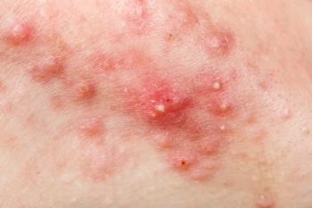
Physical examination revealed, a lesion on the right popliteal fossa described as a 4 cm erythematous plaque with areas of thick scale.
Following referral to a dermatologist, a skin biopsy was recommended and she underwent a shave biopsy, and pathology results showed proven BD, which represents SCC in situ that had extended to peripheral margins. To prevent recurrence, she underwent more extensive excision to ensure clear/negative margins and will continue following up with dermatologists for surveillance skin exams.
Discussion
BD is a type of in-situ intraepidermal SCC that has a low risk of spreading to other parts of the body.12 Both the skin and the mucosa may be affected. Around 1.42 cases per 1000 people are reported.13 It is uncommon in people with type V Fitzpatrick skin.14 The elderly are typically affected by BD, which mostly occurs in sunlight-exposed places. On the other hand, it may occur in places that are not exposed to the sun, such as the genitalia. In our case report, we presented a case of a woman aged 75 years who initially received a misdiagnosis of fungal skin infection but would not respond to an anti-fungal regimen, which highlights the challenges associated with early and accurate diagnosis in the management of BD, especially when presentation is atypical.
In their study of 1001 patients with BD, Kossard and Rosen discovered that the head and neck (44%), lower extremities (29.8%), upper extremities (19.8%), and trunk (6.5%) were the most frequently affected areas.15 In men, it is common in the anterior trunk, neck, and bald scalp. However, women are more prone to be affected in the lower legs and cheeks. Rarely, the external auditory canal, eyelid, conjunctiva, nail bed, umbilicus, lips, palms, soles, and nipple are affected.16,17 There have been few reports on the occurrence of BD on the lower limbs worldwide. Our patient, who presented with the persistent rash on the posterior aspect of her right knee, was one of the few reported cases with lower extremity BD.
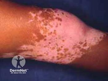
Clinically, BD manifests as a clearly demarcated single plaque that is crusty, scaly and sometimes erythematous.12 This particular form of morphology is frequently mistaken for discoid lupus erythematosus, actinic keratosis, nummular eczema, bowenoid papulosis, and psoriasis. Verrucous, hyperkeratotic, atrophic, and pigmented forms are other unusual clinical abnormalities.18 The pigmented variety among them only occurs in approximately 2% of people.9,18,19 There is only one single case report by Gahalaut et al with widespread pigmented BD.20 Widespread distribution, when it occurs, could be associated with exposure to arsenic. Generally, BD develops on skin that has been harmed by radiation, arsenic exposure, infection with the human papillomavirus, prior radiation, immunosuppression, trauma, and genetic factors.21 Though, in literature, it has never been documented that immunocompetent persons have developed extensive lesions.
In this index case, the patient presented with solitary lesion of a persistent rash area on the posterior aspect of her right knee, lasting for 15 years, highlighting the chronic nature of BD.
Patients frequently have a well-defined, erythematous, scaly patch or plaque that is asymptomatic, slowly growing, and slowly increasing. Anywhere on the mucocutaneous surface may experience it. BD frequently takes longer to diagnose because the lesion is asymptomatic; early skin changes may be subtle and resemble those of many other diseases, including psoriasis, nummular eczema, seborrheic keratosis, Paget disease, superficial basal cell carcinoma, actinic keratosis, and tinea corporis.21,22 In line with this, our patient had this rash for as long as 15 years. The rash was characterized by crusting, itching, and pain, although there was no associated bleeding, thus it was initially misdiagnosed as a fungal infection and the patient was prescribed topical ketoconazole.
A skin biopsy, which can be performed using a punch or shave biopsy, is the gold standard for the diagnosis of SCC in situ.23 A dermatopathologist should receive biopsy samples from the lesion and the tissues around it for adequate pathologic evaluation. The diagnosis of SCC in situ depends heavily on microscopic inspection. It takes into account the depth, differentiation, and invasion characteristics of the biopsy samples. It should be noted that samples of normal tissue are typically included in biopsy specimens, which can be utilized to identify differences. Additionally, a full thickness biopsy is required to accurately assess the depth of the lesion and the degree of invasion. The epidermis exhibits hyperkeratosis and parakeratosis histopathologically.
Common signs include prominent acanthosis, rete ridge lengthening, and thickening. These transform into keratinocyte cells that are exceedingly unusual. The abnormal cells exhibit mitotic activity, pleomorphism, and larger nuclei and cover the whole thickness of the epidermis. Hair follicle involvement is seen in several situations. The cells of the upper epidermis may vacuolate and have an abundant and highly eosinophilic cytoplasm.24,25
The most common form of therapy is local excision of the lesion. Other treatment options for early lesions include the use of topical creams such as imiquimod(a topical immune response modifier), or 5-fluorouracil (topical antineoplastic agent). X-ray or Grenz-ray radiation therapy can also be applied for patients with multiple lesions. Photodynamic therapy is another treatment option that has shown great success in the management of BD. A light source is then used to stimulate the injected agent, and this triggers tumor destruction.26,27 Mohs’ surgery has been the most commonly used modality in both the UK and Australia.28
There is a role for the use of topical Nystatin-Triamcinolone cream for symptom relief. Nystatin-Triamcinolone is a combination medication that combines an antifungal agent (nystatin) with a corticosteroid (triamcinolone). The dual action of this medication addresses both the inflammatory component of the rash and any potential fungal involvement, aligning with the suspected fungal infection mentioned during the patient’s previous consultation. This approach is consistent with the current management strategies for BD, focusing on symptom relief.
BD, while non-invasive, carries the potential to progress to invasive SCC if left untreated. As such, prompt intervention is necessary to prevent malignant transformation.
Prompt referral for dermatology assessment is key for quick diagnosis. Dermatologists possess specialized expertise in diagnosing and managing skin conditions, including skin cancers. The referral facilitates ongoing and comprehensive management, ensuring that the patient receives expert guidance and close monitoring. This multidisciplinary approach optimizes the patient’s chances of favorable outcomes.
BD is typically not immediately life-threatening and curable when properly managed. The majority of studies report a risk of progression to invasive SCC at 3% to 5%. According to a retrospective case series with multiple potential biases, of those that become invasive SCC, one third may metastasize. The risk of invasive carcinoma is estimated to be higher for genital BD or erythroplasia of Queyrat at 10%.29 There are rare case reports of self-resolving BD.30 Similarly, our patient presented with the lesions that had extended to peripheral margins. After surgical excision, if negative margins are not achieved, there is a higher risk of recurrence,thus the goal is to achieve clear margins.31
Conclusion
BD lesions can have an atypical presentation associated with rash and pain, in contrast to the typical presentation as a slowly enlarging erythematous to pink, scaly patch or plaque with irregular borders. This case highlights the challenges in the early diagnosis and management of BD in patients with atypical presentation, as our patient had a persistent rash for 15 years, which was initially misdiagnosed as a fungal infection.
Early recognition and management of BD is key because of its potential to progress into invasive SCC. Despite initial misdiagnosis, the patient’s lesion was accurately identified as Bowen’s disease through histopathological analysis. Further follow-up with dermatologists will ensure the continued monitoring and surveillance for treatment of the patient’s condition. Increased awareness of these atypical dermatologic manifestations will aid in the recognition and diagnosis of BD and contribute to our understanding of the underlying genetic and molecular pathways involved.
Veronica Nwagwu, MD, CWSP, is a certified wound specialist physician and clinical assistant professor in the Department of Geriatric and Palliative Medicine at Michigan Medicine in Ann Arbor, Michigan.
Nikhil Kumar Kotla, MD, is a resident in the Department of Internal Medicine at Trinity Health Oakland Hospital in Pontiac, Michigan.
Jerome Okudo, MD, MPH, is a researcher in the Department of Public Health at the University of Texas Health Science Center at Houston.
References
- Giuffrida R, Conforti C, Resende FS, et al. Clinical and dermoscopic features of genital pigmented Bowen disease. Clin Exp Dermatol. 2018 Oct;43(7):813-816.
- Bath-Hextall FJ, Matin RN, Wilkinson D, Leonardi-Bee J. Interventions for cutaneous Bowen’s disease. Cochrane Database Syst Rev. 2013 Jun 24;2013(6):CD007281.
- Costa C, Villani A, Russo D, Cappello M, Salvatores GD, Scalvenzi M. Squamous cell carcinomas in two cases of nail lichen planus: is there a real association? Dermatol Ther (Heidelb). 2018 Sep;8(3):491-494.
- Kovacs A, Yonemoto K, Katsuoka K, Nishiyama S, Harhai I. Bowen’s disease: statistical study of a 10 year period. J Dermatol. 1996 Apr. 23(4):267-74.
- Zhan W, Liao X, Tian T, Zhang RY, Li P, Wu XP, et al. Perianal multiple Bowen’s disease: a case report. Int J Clin Exp Pathol. 2015. 8 (11):15039-41.
- Chute CG, Chuang TY, Bergstralh EJ, Su WP. The subsequent risk of internal cancer with Bowen’s disease. A population-based study. JAMA. 1991 Aug 14. 266(6):816-9.
- Reizner GT, Chuang TY, Elpern DJ, Stone JL, Farmer ER. Bowen’s disease (squamous cell carcinoma in situ) in Kauai, Hawaii. A population-based incidence report. J Am Acad Dermatol. 1994 Oct. 31(4):596-600.
- Jung SE, Kim SK, Kim YC. Photodynamic therapy in Bowen disease of the first web space of the hand. Ann Dermatol. 2015 Feb. 27 (1):76-8.
- Duncan KO, Geisse JK, Leffell DJ. Epithelial precancerous lesions. In: Wolff K, Goldsmith LA, Katz SI, Gilchrest BA, Paller BS, Leffell DJ, editors. Fitzpatrick's Dermatology in General Medicine, 7th ed. New York: Mcgraw-Hill; 2008. p. 1007-27.
- Cox NH, Eedy DJ, Morton CA. Guidelines for management of Bowen’s disease: 2006 update. Br J Dermatol. 2007;156:11-21.
- Warshauer E, Warshauer BL. Clearance of basal cell and superficial squamous cell carcinomas after imiquimod therapy. J Drugs Dermatol. 2008;7:447-51.
- Singh S, Khaitan BK, Sharma MC, Seenu V, Kumawat M, Chatterjee P. Bowen’s disease on finger: a diagnostic and therapeutic challenge. Indian J Dermatol Venereol Leprol. 2013;79:227‑30.
- Neubert T, Lehmann P. Bowen’s disease‑a review of newer treatment options. Ther Clin Risk Manag. 2008;4:1085‑95.
- Quinn AG, Perkins W. Non melanoma skin cancer and otherepidermal skin tumours. In: Burns DA, Breathnach SM, Cox NH, Griffiths CE, editors. Rook’s Textbook of Dermatology. 8th ed.. Singapore: Wiley‑Blackwell; 2010. p. 32‑4.
- Kossard S, Rosen R. Cutaneous Bowen’s disease. An analysis of 1001 cases according to age, sex, and site. J Am Acad Dermatol. 1992;27:406–10.
- Neagu TP, Ţigliş M, Botezatu D, et al. Clinical, histological and therapeutic features of Bowen’s disease. Rom J Morphol Embryol. 2017;58:33–40.
- Calonje JE, Brenn T, Lazar A, Billings S. 5th ed. Elsevier; 2019. McKee’s pathology of the skin: with clinical correlation.
- Yahya H, Mohammed A. Bowen’s disease: report of a case in a Nigerian man. West Afr J Med. 2005;24:350‑1.
- Papageorgiou PP, Koumarianou AA, Chu AC. Pigmented Bowen’s disease. Br J Dermatol. 1998;138:515‑8.
- Ragi G, Turner MS, Klein LE, Stoll HL Jr., Pigmented Bowen’s disease and review of 420 Bowen’s disease lesions. J Dermatol Surg Oncol. 1988;14:765‑9.
- Gahalaut P, Rastogi MK, Mishra N, Chauhan S. Multiple pigmented Bowen’s disease: a diagnostic and therapeutic dilemma. Case Rep Oncol Med. 2012;2012:342030.
- Neubert T, Lehmann P. Bowen’s disease‑a review of newer treatment options. Ther Clin Risk Manag. 2008;4:1085‑95.
- Boer-Auer A, Jones M, Lyasnichaya OV. Cytokeratin 10-negative nested pattern enables sure distinction of clonal seborrheic keratosis from pagetoid Bowen’s disease. J Cutan Pathol. 2012 Feb. 39(2):225-33.
- Terada T. Pigmented Bowen disease arising in pigmented reticulated seborrheic keratosis. Int J Clin Oncol. 2010 Dec. 15(6):608-10.
- Palaniappan V, Karthikeyan K. Bowen’s Disease. Indian Dermatol Online J. 2022;13(2):177-189.
- Majores M, Bierhoff E. Actinic keratosis, Bowen’s disease, keratoacanthoma and squamous cell carcinoma of the skin. Pathologe. 2015 Feb;36(1):16-29.
- Zalaudek I, Argenziano G. Dermoscopy of actinic keratosis, intraepidermal carcinoma and squamous cell carcinoma. Curr Probl Dermatol. 2015;46:70-6.
- Lazarevic D, Ramelyte E, Dummer R, Imhof L. Radiotherapy in periocular cutaneous malignancies: a retrospective study. Dermatology. 2019;235(3):234-239.
- Shimizu A, Kuriyama Y, Hasegawa M, Tamura A, Ishikawa O. Nail squamous cell carcinoma: a hidden high-risk human papillomavirus reservoir for sexually transmitted infections. J Am Acad Dermatol. 2019 Dec;81(6):1358-1370.
- Morley GL, Matthews JH, Verpetinske I, et al. A comparative study examining the management of Bowen’s disease in the United Kingdom and Australia. Dermatol Res Pract. 2015;2015:5.
- Sharma A, Birnie AJ, Bordea C, et al. British Association of Dermatologists guidelines for the management of people with cutaneous squamous cell carcinoma in situ (Bowen disease) 2022. Br J Dermatol. 2023 Feb 10. 188 (2):186-194.
Newsletter
Like what you’re reading? Subscribe to Dermatology Times for weekly updates on therapies, innovations, and real-world practice tips.
Advertisement
Related Content
Advertisement


Latest CME


Advertisement
Advertisement
Trending on Dermatology Times
1
FDA Accepts Galderma’s BLA Resubmission for Liquid Neuromodulator RelabotulinumtoxinA
2
AbbVie Files for Vitiligo Indication, Putting Systemic Therapy Under Regulatory Review
3
Icotrokinra Shows Superior Efficacy Over Advanced Oral Therapies in New Psoriasis Meta-Analysis
4
Introducing Dermatology Times NP/PA Connect
5